
The Human Digestive System
TITAN MILLER
This report is about the digestive system and what role it plays in the human body. This will explain about all the parts of the digestive system and how they work. Also this will track a piece of food through the system and show how the food you eat is broken down so that it can be used and stored by your body. If it were not for your digestive system your body would not be able to extract the necessary nutrients and minerals from your food, which are needed to keep you alive.
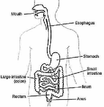
Digestion is the process where the body breaks down the food you eat so your body can use the nutrients to build up and nourish cells. It also provides energy so that you can function (Anon. 5, 2001). Your digestive system is composed of several hollow organs. These organs connect your mouth to your anus (Anon. 2, 1999). Your digestive system is about 30 feet long from the mouth to the anus (Fusco, 1998). The digestive system consists of the mouth, esophagus, stomach, small intestine, large intestine, rectum and anus. The digestive system converts the food into a form that can be used by the trillions of cells in your body (Paxton, McAndrew, 2001). There are some organs that are not part of the system but aid the process of digestion; they include the tongue, glands in the mouth that make saliva, pancreas, liver and gallbladder. Without your digestive system none of your cells would be able to acquire the energy needed to continue functioning (Anon. 4, 2001).
Digestion begins in the mouth. Food enters the digestive system via the mouth or oral cavity (Paxton, McAndrew, 2001). The mouth can reduce the size of food by grinding it with the thirty-two teeth located in the mouth. There are three types of teeth they are incisors, canines, and molars. Incisors are the teeth that you use to bite off food. They are sharp and have a straight edge to make it easy to cut food (Wortel, 2001). The canines aid the incisors in biting into food. They also tear off the food (Wortel, 2001). Molars are used to grind food (Wortel, 2001). The food physically touching nerve endings in the palate and tongue stimulate blood to circulate through the digestive system, and also increases the amount of digestive enzymes secreted such as saliva. The foods texture and temperature also contribute to this (Anon. 6, 2001). While the food is being ground in the mouth, saliva from the salivary glands is mixed into the food. When food hits saliva it mixes and causes the food particles to bind together. The mouth reducing food size and saliva mixing with the food is the beginning of the digestive system (Anon. 4, 2001).
After the food has been thoroughly chewed and saturated with saliva it is ready to leave the mouth. It does this by passing first through the pharynx located in the back of the throat and then it enters the esophagus. The pharynx is a cavity at the back of your throat that leads to the stomach (Anon. 4, 2001). Swallowing is a very complicated process that requires the tongue, soft palate, pharynx and esophagus all to engage in a sequential action just to swallow food or drink (Paxton, McAndrew, 2001). Although we are able to start swallowing by choice, once the swallowing begins, it becomes involuntary and proceeds under the control of the nerves (Anon. 2, 1999). The esophagus is the major passageway from the oral cavity to the stomach (Anon4, 2001). The esophagus is a tube that starts at the end of the pharynx and enters the stomach. It is about one quarter meter long (Anon. 4, 2001).
The stomach is a C shaped organ located on the left side of the abdominal cavity (Paxton, McAndrew, 2001). Food enters the stomach from the top by means of the esophagus. A large percentage of digestion happens in the stomach. Several times per minute, automatically controlled rippling waves pass through your stomach that mixes gastric juices, food and digestive enzymes that break the food down further (Anon. 6, 2001). The stomach has three main jobs to do. Its first job is to store all of the food and liquid that you swallowed. In order for the food to enter the stomach the muscle at the top of the stomach must relax which opens the valve at the top so the food can enter the stomach. The stomach's next job is to mix the food, liquid, and digestive juices produced by the stomach. To mix the food the stomach must use the muscles in its lower half to churn the contents. The stomach's last task is to slowly empty its contents into the duodenum (Anon. 2, 1999). One of the stomach's most important functions is its ability to secrete gastric juices. The secretion of gastric juices from the stomach is controlled by the neural and hormonal systems (Anon. 4, 2001). The inner most lining of the stomach is a thick mucus membrane and it has many small holes in it. The holes are called gastric pits. The gastric pits are located at the end of the gastric glands (Anon. 4, 2001). Inside of the gastric glands there are several different types of cells these secondary cells are mucous cells, chief cells, and parietal cells. Mucous cells are located near the openings of the gastric pits. The chief cells and parietal cells are found further down inside of the glands (Anon. 4, 2001). The mucous secreted by the mucous glands acts as a protective coating against pepsin and accidentally swallowed sharp objects (Anon. 4, 2001). Pepsin is a substance that is secreted by chief cells as an inactive enzyme called pepsinogen. When pepsinogen interacts with the hydrochloric acid produced by parietal cells, it changes into pepsin (Anon4, 2001). Pepsin functions as a protein splitting enzyme (Anon4, 2001). Pepsin is most effective in an acid-filled environment. The high concentration of hydrochloric acid provides an excellent environment for pepsin to begin the digestion of proteins (Anon4, 2001). Parietal cells release hydrochloric acid that increases the efficiency of pepsin. When pepsin, mucus and hydrochloric acid are combined it is called gastric juices. Once food has been mixed with gastric juices it becomes a semi-fluid paste called chyme. The food now passes through the bottom valve of the stomach into the small intestine.
After the chyme exits the stomach it enters the duodenum. The duodenum is the first part of the small intestine (Paxton, McAndrew, 2001). The duodenum is C shaped and curves around the head of the pancreas. The small intestine is composed of two other parts besides the duodenum. They are the jejunum and the ileum. The jejunum is on average a length of eight feet long and the ileum is 12 feet long. The small intestine is tubular in shape and has many coils and loops. It occupies a large percentage of the abdomen. Two organs help the small intestine process food. They are the pancreas and the liver. They produce juices that aid digestion. The pancreas produces a juice that contains a wide array of enzymes to break down the carbohydrates, fat, and protein in our food (Anon2, 1999). The liver produces another digestive juice called bile. The bile is stored between meals in the gallbladder. At mealtime it is squeezed out of the gallbladder into the bile ducts to reach the intestine and mix with the fat in our food. The bile acids dissolve the fat into watery contents of the intestine (Anon2, 1999). The bile stored in the gallbladder is composed of bile salts, bile pigments, cholesterol, and various electrolytes. Bile goes into the duodenum only when the gallbladder is stimulated to contract by the hormone cholecystokinin (Anon4, 2001). Bile salts affect the fat-soluble vitamins A, D, E, and K. Fat molecules naturally clump together. When they do, they form fat globules. The masses of fat formed are difficult to digest, and this is where bile salts come in. Bile salts cause fat globules to break up. This makes it easier for fat splitting enzymes to digest the fat molecules. The process of breaking up fat globules is called emulsification (Anon4, 2001). To maximize absorption, the food molecules must come in contact with a large number of specialized cells. To accomplish this, each square inch of the intestinal wall is covered with about ten billion microscopic hair like projections called microvilli, which absorb nutrients into your blood stream (Anon4, 2001). If your intestinal interior were smooth, it would present only about six square feet of absorptive surface. Instead, because of these microvilli, it presents about 4,000 square feet (Anon4, 2001). At the base of the villi there are glands that secrete a large amount of a watery fluid. The watery fluid secreted by the base of the villi acts as a medium in which digestive products can move. Once the digestive products are in the watery fluid, they can then be moved into the villi. Basically, the watery fluids are a vehicle that allows villi to acquire the needed digestive products (Anon4, 2001). Food is propelled along the small intestine with a method called peristalsis. The action of peristalsis looks like an ocean wave moving through the muscle. The muscle of the organ produces a narrowing and then propels the narrowed portion slowly down the length of the organ. These waves of narrowing push the food and fluid in front of them through each hollow organ (Anon2, 1990).
The next organ in the digestive system is the large intestine. The large intestine has four basic functions: the reabsorption of water, the reabsorption of electrolytes, the formation of feces, and the storage of feces (Anon4, 2001). But, when compared to the small intestine, the large intestine plays a very small part in the digestive process (Anon4, 2001). The large intestine extracts water from the waste material and what remains moves via peristalsis through the five feet of the large intestine to the rectum for eventual elimination in the form of a bowel movement. The large intestine begins in the cecum. The cecum continues as the ascending colon upward along your right side. Just under the liver, it makes a sharp turn. The colon continues across the upper abdomen as the transverse colon, only to make another sharp downturn. The colon continues straight down your left side as the descending colon and then into an S-shaped segment called the sigmoid colon (Fusco, 1999). The large intestine continues as the rectum. The rectum is the straight part at the end of the large intestine. At the end of the rectum is the anal canal. The anal canal is the very end of the digestive system. Just as at the beginning of the digestive process the chewing of food in the mouth is controlled by our conscious actions, voluntary control returns at this end of the digestive tract, during elimination (Anon6, 2001).
To explain the digestive process I will be following a quarter pound hamburger and 12 ounces of pop through the digestive system. I will try to explain what happens to the food in the various organs of the digestive tract.
You sit down and you are presented with a nice quarter pound hamburger and a 12 ounces cup of your favorite pop. You open the wrapper on the hamburger and as you inhale through you nose the smell stimulates your salivary glands to produce saliva. You then open your mouth and take a bite. As soon as the food touches your tongue your salivary glands produce even more saliva to break down starch in the food and to make the food bind together. Once the food is firmly in your mouth you begin to chew with your 32 teeth to physically break down the food into smaller bits to make it easier to swallow.
After you determine that the food has been ground small enough to swallow your body goes into the buccal phase, which is the only voluntary stage of digestion. Your tongue forces food into the pharynx. After this the process is reflex. The tongue blocks the mouth, soft palate closes off the nose and the larynx rises so that the epiglottis closes off the trachea (Paxton, McAndrew, 2001). The bite of food now enters the esophagus and is propelled down by peristalsis aided by gravity. Peristalsis is the process where the organ narrows down at the top or beginning and then the narrow part is "pushed" along the organ. Peristalsis is more drastic in the esophagus than it is in an organ like the small intestine.
After the food leaves the esophagus it enters the stomach where it stays for several minutes to one hour or more depending on how large of a meal you ate. By the time the bite has gotten to the stomach you feel like a drink so you take a sip of your beverage. You do not need to chew a liquid though because it can go down the esophagus without any trouble. When you swallow a liquid as it goes down the esophagus although the esophagus still uses peristalsis the gravity causes the liquid to fall at a greater rate than the peristalsis travels.
In ten minutes or so you are completely done with your meal and decide to sit down and watch television. As you sit there a rumbling sound is coming from your abdomen. This noise is your stomach mixing your food with gastric juices. The gastric juices including pepsin and hydrochloric acid break down your food into a paste called chyme.
Your television program has gone off and the great taste of the hamburger has dissipated. You go brush your teeth and go to bed. Your stomach has been gradually letting food seep out of its bottom valve into the duodenum. The duodenum is the first part of the small intestine. When the food is in the small intestine bile secretes into the small intestine from the gallbladder. The gallbladder is actually only the storage area for the bile; the pancreas is what actually produces the bile. Bile contains bile acids, which are critical for digestion and absorption of fats and fat-soluble vitamins in the small intestine (Anon7, 1999). Adult humans produce 400 to 800 ml of bile daily (Anon7, 1999). Free cholesterol is virtually insoluble in aqueous solutions, but in bile, it is made soluble by bile acids and lipids like lethicin (Anon7, 1999). Large amounts of bile acids are secreted into the intestine every day, but only relatively small quantities are lost from the body. This is because approximately 95% of the bile acids delivered to the duodenum are absorbed back into the blood within the ileum (Anon7, 1999). Now the bile has broken down most of the cholesterol in the hamburger, which makes the paste thinner and more fluid. The wall of the jejunum is lined with villi. Villi increase the surface area of the small intestine. Inside each villi is a capillary, which is the smallest type of blood vessel in the body. The nutrients soak through the villi's wall and into the capillary to be used throughout the body. While in the jejunum, at the base of the villi there are small glands that produce large amounts of watery fluid that helps the chyme move along the small intestine and greatly improves the villi's ability to absorb nutrients. By the time the hamburger has reached the end of the small intestine it is very watery and has most of its useful material removed. The chyme moves along the small intestine by peristalsis, but it is much weaker in the small intestine than the esophagus. As the chyme leaves the jejunum and enters the ileum digestion is basically complete. It has been around eight hours since you ate the hamburger and pop and the chyme is now entering the large intestine.
The large intestine removes water from the chyme and absorbs electrolytes that were released into the chyme during digestion. It also forms and stores feces. The chyme first enters the cecum, which is the first part of the large intestine. Then it enters the colon, which is the ribbed portion of the large intestine. As it travels the length of the colon water and liquids are removed leaving only the solid portions. The large intestine secretes mucous which holds the feces together. It will take from one to two days for the feces to work its way along the large intestine where it eventually enters the rectum. The rectum is where the large intestine straightens out to leave the body. The very last part of the large intestine and the digestive system is the anal canal. The anal canal is where the feces exits the body. This is how the food you eat is broken down to supply you with energy to function.
This report was about the digestive
system and what role it plays in the body. It explained how all of the organs
in the digestive system work together to convert the food you eat into energy
that powers your body.

Literature Cited
Anonymous2. 1999. "Your Digestive System and How It Works." [Online] 2 November, 2001. NIH publication No. 99-2681 December 1998.
http://www.niddk.nih.gov/health/digest/pubs/digesyst/newdiges.htm
Anonymous4. 2001. "The Digestive System." [Online] 2 November, 2001.
http://www.msms.doe.k12.ms.us/teachers/wodom/anatomy-old/digestive/digestive.html
Anonymous5. 2001. "The Digestive System - an Overview." [Online] 6 November, 2001. ViaHealth.
http://www.viahealth.org/disease/digestive/howworks.htm
Anonymous6. "The Human Body and How It works." [Online] 10 November, 2001. Columbia University College of P&S Complete Home Medical Guide.
http://cpmcnet.Columbia.edu/texts/guide/hmg04_0001.html
Anonymous7. 1999. "Secretion of Bile and the Role of Bile Acids in Digestion." [Online] 10 November, 2001.
http://arbl.cvmbs.colostate.edu/hbooks/pathphys/digestion/liver/bile.html
Fusco, Robert D. M.D. 1998. "How Does Your Digestive System Works?" [Online] 2 November, 2001. Crohn's & Colitis Foundation of America, Inc. Copyright ã 1996-2001.
http://www.ccfa.org/weekly/wkly911.htm
McAndrew, Terry and Paxton, Steve. "Introductory Anatomy: Digestive System." [Online] 1 November, 2001. Center for Human Biology.
http://www.leeds.ac.uk/chb/lectures/anatomy8.html
Wortel, John P. "Teeth." [Online] 13, November, 2001. Discovery Channel School.
http://school.discovery.com/homeworkhelp/worldbook/atozscience/t/549500.html